-
 Application Areas
-
Application Areas
-
 Forensic medicine
 Forensic medicine plays a critical role in ensuring justice by providing scientific data to the legal system. The examination and analysis of criminal evidence is the specialty of forensic laboratories.
 Experimental Animals
 Advanced Instrumentation Solutions for Laboratory Animals: Scientific Ethical Research Experimental animals play an indispensable role in the development of new drugs, treatments, vaccines and surgical techniques, in understanding biological processes and in elucidating disease mechanisms. This critical role also requires laboratory animal laboratories to operate at the highest scientific and ethical standards. Ensuring animal welfare, data reliability and research reproducibility are the main priorities of these laboratories. Our company provides an ethical and reliable basis for your scientific research by providing high-tech laboratory instruments and integrated solutions required by laboratories in this sensitive and regulated field.
 Cell Theraphy-GMP/GLP
 Advanced Instrumentation Solutions for Cell Therapy and GMP/GLP Laboratories: From Safety to Treatment Cell and gene therapies offer a wide range of innovative treatment options, from cancer to genetic diseases, with autologous (from the patient’s own cells) or allogeneic (from donor cells) approaches. The development and production of these therapies require the highest standards of safety, efficacy and quality for human health. Cell therapy laboratories must strictly adhere to the principles of Good Manufacturing Practice (GMP) and Good Laboratory Practice (GLP) throughout the process, from research and development to clinical application. Labor Ildam supports the delivery of safe and effective cell therapies to patients by providing high-tech, validated and compliant laboratory instruments and integrated solutions required by laboratories in this highly regulated and technically challenging field.
 Biobank RNA&DNA&Tissue
 Advanced Instrumentation Solutions for Biobank (RNA, DNA, Tissue) Laboratories: From Sample Integrity to Scientific Discovery Biobanks are invaluable resources that form the basis of modern biomedical research and personalized medicine. Access to high-quality biological samples (RNA, DNA, tissue, blood, plasma, etc.) is critical for a wide range of purposes, from understanding the causes of diseases to developing new diagnostic methods and treatments. The collection, processing, long-term storage and distribution of these samples require the expertise of biobank laboratories that maintain sample integrity, traceability and data security at the highest level. Our company supports the protection of your valuable biological resources and their transformation into scientific discoveries by providing high-tech, reliable and automated laboratory instruments and integrated solutions required by laboratories in this highly sensitive and regulated area.
 BSL 1 2 3 4 Laboratories
 Advanced Instrument Solutions for BSL 1, 2, 3 and 4 Laboratories: Maximum Safety, Minimum Risk Biological research and diagnostic studies are essential for human health and public safety. However, potential biological risks that may be encountered during these studies require the protection of laboratories and employees. Biosafety Level (BSL) laboratories are specially designed and equipped facilities classified according to the degree of danger of the microorganisms studied (Risk Group 1 to 4). Each level from BSL-1 to BSL-4 offers an increasing level of protection and isolation. Our company offers state-of-the-art laboratory instruments and integrated solutions that meet these stringent standards, combine safety and efficiency, and help you maximize the safety of your research and operations when working with biological hazards.
 Bone Marrow Transplantation
 Advanced Instrumentation Solutions for Bone Marrow Transplantation Laboratories: Assurance of Promising Therapies Bone marrow transplantation is one of the most effective treatments in modern medicine for life-threatening blood diseases, genetic disorders and some types of cancer. This complex and delicate process requires the highest standards of sterility, precision and quality control at every stage, from the collection of stem cells from the donor to their processing, storage and transplantation to the patient. Bone marrow transplantation laboratories play a critical role in the success of these vital treatments. Our company provides state-of-the-art, validated and highly reliable laboratory instruments and integrated solutions that laboratories in this specialized and regulated field need, thus ensuring promising therapies for patients. Key Challenges in Bone Marrow Transplantation Laboratories and Our Solutions Bone marrow transplantation laboratories must cope with the special challenges of working with living cells (cell viability, risk of contamination) and strict regulatory requirements (traceability, data integrity, GMP/GLP compliance)
 Cord Blood Banking
 Advanced Device Solutions for Cord Blood Banking Laboratories: Investing in the Health of the Future Cord blood banking is a rapidly developing field that enables the storage of potentially life-saving stem cells for future treatment needs. This valuable biological material obtained from the umbilical cords of newborn babies offers an alternative source for the treatment of many diseases, especially hematological and genetic diseases. Cord blood banking laboratories in this field require absolute sterility, high precision and strict regulatory compliance (GMP/GTP) at every stage from cord blood collection to processing, from quality control to cryopreservation. Our company supports you in preserving the integrity of your valuable stem cell products and paving the way for future treatments by providing state-of-the-art, validated and highly reliable laboratory devices and integrated solutions required by laboratories in this special and meticulous field.
 IVF-In Vitro Fertilization
 Advanced Device Solutions for IVF Laboratories: Scientific Support for the Formation of Future Families In vitro fertilization (IVF), or in vitro fertilization treatment, is one of the most miraculous areas of medicine, offering millions of couples struggling with infertility the opportunity to realize their dreams of parenthood. The success of this treatment is directly dependent on the meticulous and delicate processes carried out in IVF laboratories. From the collection of egg and sperm cells to every stage of embryo development, the highest sterility, optimal environmental conditions and state-of-the-art laboratory devices are of critical importance at every step. Our company provides scientific and technological support for the formation of future families by offering the highly sensitive, reliable and innovative solutions required by laboratories in this special and sensitive field.
 Blood Banking
 Advanced Device Solutions for Blood Banking Laboratories: The Assurance of Life-Saving Drops Blood is an indispensable resource for human life, and blood banking is a vital service that ensures patients have access to the blood and blood products they need during surgical operations, traumas, chronic diseases or cancer treatments. This critical process requires the highest standards of safety, precision and quality control at every stage, from blood collection to virus screenings, blood group tests, component separation and long-term storage. Blood banking laboratories are the heart of transfusion medicine and ensure that every drop of blood reaches the patient safely. Our company provides the assurance of life-saving transfusion services by providing state-of-the-art, validated and highly reliable laboratory devices and integrated solutions required by laboratories in this special and regulated field.
 Artificial Insemination/Veterinary Medicine
 Artificial Insemination and Advanced Instrumentation Solutions for Veterinary Laboratories: Healthy Animals, Efficient Production Animal health and efficient animal husbandry are of great importance for both food safety and biodiversity protection. While artificial insemination forms the basis of modern animal husbandry by accelerating genetic improvement and increasing reproductive efficiency, veterinary laboratories play a key role in the diagnosis, prevention and treatment of animal diseases, vaccine development and general animal welfare. In this dynamic and diverse sector, state-of-the-art laboratory instruments and precise analytical methods are needed for accurate diagnosis and effective solutions. Our company provides scientific support for both animal health and sustainable animal production by offering the high-precision, reliable and innovative solutions required by laboratories in this special field.
 Cell Culture
 Advanced Instrumentation Solutions for Cell Culture Labs: A Cornerstone in Life Sciences Cell culture is a cornerstone of modern life sciences and biotechnology. From cancer research to drug discovery, vaccine production to regenerative medicine, the ability to keep cells alive, grow, and manipulate them in the laboratory is essential for many fields. These delicate processes require the expertise of cell culture laboratories and state-of-the-art laboratory instruments that provide controlled, sterile, and optimized conditions that best mimic the cells’ natural environment. Your company can help you increase the accuracy of your research and accelerate scientific discovery by providing the high-precision, reliable, and efficient solutions that laboratories in this critical field need.
 Microbiology
 Advanced Instrumentation Solutions for Microbiology Laboratories: Exploring the Microscopic World Microbiology provides a wide range of solutions, from human health to environmental protection, from food safety to industrial processes, by revealing the secrets of the invisible world. The study of bacteria, viruses, fungi and other microorganisms requires the indispensable expertise and state-of-the-art laboratory equipment of microbiology laboratories in every field, from disease diagnosis to the development of new antibiotics. The reliability and accuracy of every data obtained is of critical importance for both scientific progress and public health. Your company contributes to the success of your microbiological research and operations by providing the precise, reliable and high-performance analytical solutions required by laboratories in this dynamic and demanding field
 Equipment for Clinical Research
 Advanced Instrumentation Solutions for Clinical Research Laboratories: Hope from Science to Patients Clinical research is a critical bridge that transforms scientific discoveries into tangible treatments for improving human health. From testing the efficacy of new drug candidates to verifying the safety of existing treatments, the meticulous and precise work of clinical research laboratories is essential at every step. These laboratories face a wide range of challenges, from patient sample collection to complex analyses, from data integrity to compliance with strict regulatory standards (Good Clinical Practice - GCP). By providing state-of-the-art, validated and highly reliable laboratory instruments and integrated solutions required by laboratories in this special and highly responsible field, your company contributes to the transformation of your scientific discoveries into safe and effective treatments.
-
-
 Sectors
-
Sectors
-
 Food
Food Comprehensive Instrument Solutions for Food Laboratories: From Safety to Quality The food industry is of vital importance for human health and economies. Access to safe and quality food for consumers is the responsibility of producers as well as the fundamental task of food laboratories that analyze these products. Developing technologies and increasing regulations constantly encourage food laboratories to use faster, more accurate and more reliable analysis methods. This is where our company offers innovative laboratory instruments and solutions that will meet all the needs of laboratory professionals in the food industry.
 Vaccine and Pharmaceutical
 Advanced Instrument Solutions for Vaccine and Pharmaceutical Laboratories: From Research to Production The vaccine and pharmaceutical industry is a dynamic field shaped by constantly evolving scientific discoveries and strict regulations. The development, production and quality control of these products, which are of vital importance to human health, require advanced laboratory equipment and precise analysis methods. Labor İldam offers innovative and reliable laboratory equipment to meet all the needs of laboratories in this critical sector, from R&D to production, from quality control to stability studies.
 Biotechnology
 Advanced Instrumentation Solutions for Biotechnology Laboratories: From Discovery to Application Biotechnology is a groundbreaking branch of science that uses living systems and biological processes to develop products and technologies. The biotechnology sector, which offers revolutionary solutions in many areas from medicine and pharmacy to agriculture, from energy production to environmental cleaning, is at the center of laboratory studies that require continuous innovation and high precision. Labor İldam helps you accelerate your research, optimize your production processes and make groundbreaking discoveries by providing state-of-the-art laboratory instruments and integrated solutions needed by biotechnology laboratories in this dynamic and rapidly growing field.
 Agricultural Research and Development (R&D)
 Advanced Instrument Solutions for Agricultural R&D Laboratories: From Efficiency to Sustainability Agricultural Research and Development (R&D) plays a vital role in meeting the increasing food needs of the world population, combating the effects of climate change, and developing sustainable agricultural practices. Modern agricultural R&D laboratories aim to increase the efficiency and quality of agricultural production by performing a wide range of analyses, from soil analysis to plant genetics, from product quality to environmental monitoring. Labor İldam provides scientific support for your agricultural research by providing high-tech laboratory instruments and integrated solutions required by laboratories in this dynamic sector.
 Electronics and Optics
 Advanced Instrumentation Solutions for Electronics and Optics Laboratories: From Precision to Innovation The electronics and optics sector is one of the cornerstones of modern technology. This sector, which finds a wide range of products and applications from semiconductors to laser systems, communication devices to imaging technologies, requires constant innovation and high precision. Electronics and optics laboratories are the heart of research, prototyping, production processes and quality control activities in this field. By providing state-of-the-art laboratory instruments and integrated solutions required by laboratories in this dynamic and demanding sector, Labor İldam accelerates your innovation processes and helps you optimize the performance of your products.
 Cosmetics
 Advanced Instrumentation Solutions for Cosmetics Laboratories: From Beauty to Safety The cosmetics industry is a multi-billion dollar global industry encompassing a wide range of products, from skin care to makeup, hair products to fragrances. As consumer interest in beauty and personal care products increases, the safety, efficacy and quality of these products are becoming increasingly important. Cosmetics laboratories play a central role in many critical processes, from the development of new formulations to raw material analysis, from quality control of finished products to stability testing. By providing state-of-the-art laboratory instruments and integrated solutions that laboratories in this dynamic and competitive industry need, Labor İldam can accelerate your innovation processes and support the success of your products in the marketplace.
 Automotive
 Advanced Instrumentation Solutions for Automotive Laboratories: From Innovation to Quality The automotive industry is one of the most complex and dynamic areas of today’s technology and engineering. The development of new generation vehicles, advances in electric vehicle technologies, autonomous driving systems and increasing environmental regulations are constantly pushing automotive laboratories to use more innovative, more precise and more reliable analytical solutions. Automotive laboratories, which are of critical importance in a wide range of areas from material characterization to fuel analysis, from emission control to battery tests, are the center of research and quality assurance that shape the future of the industry. By providing the state-of-the-art laboratory instruments and integrated solutions that laboratories in this demanding industry need, Labor İldam accelerates your innovation processes and helps you optimize the performance of your products.
-
-
 Representations
-
Representations
-
 Haier Biomedical
 Haier Biomedical was established to focus on the research and development, production and sales of low-temperature storage equipment for biomedical samples. Based on the concept of IoT, the company has become a digital scenario solution provider in the field of life science and medical innovation. Aiming to create the best user experience, the company mainly operates in the field of life science and medical innovation, and provides comprehensive digital scenario solutions covering multiple product and service categories such as smart laboratory, digital hospital, smart public health, smart blood utilization to a wide range of user groups such as hospitals, biopharmaceutical enterprises, universities and colleges, scientific research institutions, disease control centers and blood stations.
 PHC Corporation, Biomedical Division (PHCbi)
 Phcbi (Sanyo, Panasonic), which is the Turkish distributor of Labor ildam Laboratory Materials Trade Co. Ltd. Şti., was founded in 1990 as a subsidiary of PHC Holdings Corporation and aims to be a leading, trusted brand for sustainable health and biomedical product solutions that support users' efforts to improve the health and well-being of people around the world. It is a company that has been responding to the needs of pharmaceutical, biotechnology, hospital, clinical and industrial customers for over 25 years and offers a unique perspective on scientific research in general. PHCbi, which we are the Turkish distributor of, plays a critical role in product development for applications worldwide and has a reputation as a manufacturer of high-quality and innovative medical and laboratory equipment. Phcbi has established long-term relationships with leading academic and research institutes in Europe as well as leading pharmaceutical, healthcare and biotechnology companies. It is a company that sets the standard in many ways. VIP panels, Cool Safe compressors, Active Background Contamination Control and the world's first -152 C ULT freezer. The Sanyo and Panasonic brands, which have been identified with the Labor Ildam company in Turkey for many years, now continue their existence in the market under the name PHCbi.
 Esco Scientific
 World-leading manufacturer and service provider of life science instruments Esco Lifesciences is a world-leading life science company with a diversified portfolio and sales in more than 100 countries. As a manufacturer of laboratory and biopharma equipment and IVF medical devices, Esco offers tailored solutions to fit the needs of laboratories in a variety of industries.
 IC Biomedical
 Creating Greater Value for Our Customers and End Users On October 30, 2020, Milton Street Capital announced the simultaneous acquisition of Worthington Industries (formerly Taylor-Wharton CryoScience products) and the Life Sciences business of International Cryogenics, Inc. These former cryogenic biomedical equipment manufacturers later became IC Biomedical. IC Biomedical brings new life to the cryogenic equipment market by leveraging its 66-year legacy of cold chain storage and transportation technology. IC Biomedical builds the highest quality cryogenic storage and transportation systems for the global biomedical research and development, healthcare, biowarehouse, pharmaceutical, biotechnology, IVF and livestock semen markets.
 Frimed
 Frimed was founded in 1997 as a result of the twenty years of experience of its owners in the field of medical refrigeration. Continuous research and development, together with a professional and serious attitude, have enabled Frimed to design and produce highly reliable refrigerators and freezers for use in hospitals, industry, laboratories, biomedical and pharmaceutical areas and in the scientific field in general. Within a few years, our devices have become well known and are now appreciated and preferred in many countries around the world.
 Nanoentek
 Pioneer in POCT (Point Of Care Testing)! A pioneer in POCT, NanoEntek, which has the world's best microfluidic chip development and manufacturing technology, has an important Bio-MEMS technology that seamlessly integrates biotechnology with Micro-electro Mechanical Systems (MEMS), which is considered the most advanced technology of the 21st century. To realize this technology, NanoEntek's activities include R&D, manufacturing and sales of life science laboratory equipment, in vitro diagnostic medical devices and related consumables and solutions. Our company has about 100 patents related to NT-IT-BT (nanotechnology-information technology-biotechnology) convergence technology in Korea and abroad. In particular, we have the technology for an important platform called lab-on-a-chip, which integrates laboratory processes on a microfluidic chip the size of a fingernail and has made significant contributions to the fields of medical and life science engineering.
 Auguste Cryogenics
 "Auguste Cryogenics Slovakia s.r.o.", a member of the Auguste Cryogenics group with a production site in Slovakia and a distribution center in Germany, is a leader in the production technology, supply and innovation of cryogenic storage tanks. Thanks to the efforts of a team of highly professional and motivated experts, Auguste Cryogenics Slovakia s.r.o. is synonymous with reliability, quality, high level of technical expertise and "fair play". Since 2001, the production site in Košice, Slovakia has become an advanced European manufacturer and innovator with world-class quality and complete customer satisfaction as its main goal.
 Steelco
 Founded in Italy in 2001 and part of the Miele Group since 2017, Steelco S.p.A. is today one of the world's leading manufacturers of cleaning and sterilization systems for healthcare, pharmaceutical and research laboratories. A benchmark in health protection and infection control, Steelco offers its customers tailored end-to-end solutions that are increasingly efficient and reliable, automated and sustainable, maximizing safety levels, improving procedures and reducing operating costs.
 Hettich
 For over 120 years, Andreas Hettich GmbH has been a pioneer in the development and production of centrifuges and laboratory equipment used worldwide. Founded in 1904 and headquartered in Tuttlingen, Germany, our company has a rich and dynamic history. From a small workshop founded by master mechanic Andreas Hettich, we have grown into an internationally renowned organization with over 500 employees. Today, under the guiding principle of "Heritage Meets Future", we continue to operate from our headquarters in Tuttlingen, promoting innovation and setting new standards in the field of medical technology.
 Macopharma
 Macopharma is a family business with a strong sense of belonging. To protect a family and ensure the growth of everyone, we need a home with solid foundations: our values. A home also needs a roof, this is our vision, while aiming for a bright and meaningful future, we rely on our mission daily: supporting life. Our home has solid pillars that represent our roadmap: life, solutions and learning Every Macopharma employee has a place in this home This is also open to stakeholders, so together we contribute to a safer and more sustainable blood industry. Discover every section of our Macopharma Strategic Home
 Telstar
 Specializing in GMP consulting, engineering and construction projects and development of integrated process equipment, we serve companies related to Life and Health Sciences (pharmaceutical and biotechnology, healthcare, cosmetics, veterinary and food and beverage industries) as well as hospitals, laboratories and research centers. We also offer vacuum and high vacuum technologies and solutions for large scientific experiments for traditional and high-tech industries in the power and aerospace sectors. Fully integrated process solutions With over 60 years of experience in the development of complex GMP projects for the life sciences industries, from consulting, engineering and construction to integrated process equipment, we offer advanced solutions for the scientific and aerospace sectors. With global vision and fully owned expertise. As a result, our brand has emerged as a solid technology partner for the pharmaceutical and related industries, laboratories, research centers and general industry. We are recognized as one of the major international manufacturers with the capacity and knowledge to offer integrated process solutions using in-house technologies, and are valued for our role as a one-stop solution provider for aseptic production and research processes. Our customer at heart Our corporate structure is supported by a holistic vision of multidisciplinary and integrated business management. For this reason, our organization is based on a management model that prioritizes cooperation and promotes synergies between different internal operational areas covering different stages of the value chain for the customer. Knowledge and expertise in technology research, innovation, automation and product and project development are supported by a well-developed technical capacity together with a dedicated and solid professional team. Building projects and sharing knowledge, we strengthen our assets, expertise and experience by placing the customer at the center of our focus, while reinforcing our competitive value and uniqueness: offering highly competitive and complete, integrated solutions. Driving force We are a dynamic organization and our innovative structure allows us to constantly drive new innovative ideas and projects. Our dedicated and highly qualified team collaborates across the organization to deliver high-value solutions, responding to complex market demands.
 Miele Professional
 Since the founding of our company, we at Miele have believed in building a culture of innovation. We work together to deliver added value to our customers and develop the best solutions. We are in demand worldwide as a reliable partner representing top quality and expertise. With our industrial solutions, we support many sectors from nursing homes to fire brigades, hospitals to kindergartens, laboratories to laundries, and offer holistic solutions for your business.
 Afi Groups
 Laboratory equipment made in France Located in Château-Gontier, FRANCE, the French designed and manufactured AFI centrifuges are designed to meet the needs and requirements of clinical analysis and biomedical research laboratories. Offering a variety of different rotors and accessories, the user will have all the solutions for the separation and preparation of samples, whether from blood sampling in basic biological research, medical, pharmaceutical or cell culture. Available in a ventilated, refrigerated version, the AFI centrifuges are designed to be used with all tubes, bottles and flasks available on the market, as well as for the preparation of samples in microplates. The range of AFI centrifuges covers the different Clinical, Industrial and Research markets.
 Raypa
Raypa Expert manufacturer, unique design, global brand With over 50 years of experience, RAYPA has emerged as a globally recognized leader in laboratory equipment manufacturing, offering innovative solutions on an international scale. Our focus is on the development, production, marketing and after-sales service of autoclaves and food analysis equipment.
 Sherwood Scientific
 Sherwood Scientific’s history goes back over 50 years to the founding of one of our predecessor companies, Evans Electroselenium Ltd. This company was founded in the late 1930’s by Arthur Evans and was based on the use of selenium photocell applications. The Flame Photometer and CHROMA Filter Photometer series are examples of this use. Sherwood Scientific products, now manufactured entirely in Cambridge, have also been associated with the Corning and CIBA Corning brands for a number of years. Johnson Matthey is another company in the Sherwood story. Johnson Matthey is heavily involved in the precious metals sector and two of Sherwood’s products, the Fluid Bed Dryer and the Magnetic Susceptibility Balances, were developed under their auspices. Since the acquisition of these product lines, Sherwood Scientific has developed several new versions of these products. The Model 420 is a Dual Channel Flame Photometer with Internal Standard and is available for Clinical and Industrial samples. We have also developed the Model 420Cs Flame Photometer, which comes with a Cesium reference channel as an alternative to the traditional Lithium reference channel. The Model 360 is designed for ease of maintenance while allowing analysis of one element at a time. The competitively priced Model 360 comes standard with five filters for Sodium, Potassium, Lithium, Calcium and Barium. The AUTO MSB uses the same measuring principle as the classic Evans design but is 200 times more sensitive. Sherwood added Microprocessor control of air flow, inlet air temperature and drying time to the Tornado dryer purchased from Johnson Matthey and Sherwood’s latest model, the Model 501 Fluid Bed Dryer, features membrane-sealed controls to prevent particle ingress and also reduces operating noise.
 Terra Food- Tech
 Compact autoclaves for canned food and ready meals TERRA Food-Tech ® autoclaves can sterilize and pasteurize canned, glass or plastic packaged foods. Designed for cooking, sterilizing and pasteurizing packaged foods using a heart temperature probe inserted into the product sample. This allows perfect control of the entire process while preserving the organoleptic properties and minimizing possible changes in the nutritional properties of the final product.
 Snijderlabs
 Since 2008, a new generation of 'Snijders' has further driven the company's globalisation. Together with their team, Pieter and Norman Snijders have built on the foundations that were so solidly laid nearly 50 years ago. In 2013, Snijders sales activities were divided into two divisions: Laboratories and Maintenance. Snijders Maintenance offers logistics solutions for internal transport in care institutions and hospitals. Snijders Laboratories offers high-tech storage and cultivation equipment for laboratories. Snijders has always distinguished itself through short lines of communication between design and production. This formula has proven to be effective. These short lines and our own production make special, individual orders possible. Listening carefully to the needs of users has given Snijders the innovative know-how that we are currently navigating and adjusting. And of course, we also owe a debt of gratitude to all 60 employees!
 Saturix by FG Cleanwipes
 Saturix from FG Clean Wipes is a complete surface cleaning system for all surfaces cleanrooms and other controlled environments. It helps our end users reduces risk, promotes employee safety while improving operational efficiency. The system includes a focused portfolio of cleanroom wipes, cleanroom mops, innovative mop buckets and isolator cleaning tool, sterile IPA as well as other specialty products. As a trusted partner with 50+ years of cleanroom expertise, FG Clean Wipes has the experience, resources and scale to accommodate any custom or private label requirements – with the most precise, advanced and efficient solutions around the globe.
 Animal Care Systems
 Animal Care Systems unique motor-free carousel design provides the highest and most ergonomic cage density. Our microenvironments are free of noise, vibration, and ultrasound. Our patented airflow system directs and filters room air through the cages and racks directly into the building’s HVAC exhaust system. This system creates a calm, laminar airflow that provides an environment ideal for housing, experiments, and breeding. Our pie-shaped cage has been enhancing rodent life since 1997. Results obtained from Animal Care Systems cages are reliable, advantageous, and trouble-free.
 RWD Life Science
 As a world's leading manufacturer of scientific equipment company, RWD Life Science (referred as RWD), is committed to provide high-quality and cost-effective laboratory instruments to researchers, and animal medical equipment to veterinarians worldwide. The product solutions range from animal surgery and modeling, neuroscience research, animal behavior research, cell and molecular biology, microcirculation monitoring, pathological diagnosis, and animal healthcare. Since 2002, RWD, headquartered in China with a branch in the USA, has successfully provided products and services to over 100 countries and regions. We have served 6,000+ universities, 1,000+ research institutes, 2,300+ hospitals, and 11,000+ veterinary clinics, gaining constant trust and support from global customers. Looking into the future, we shall adhere to the core values of quality, integrity, responsibility and contribute to the advancement and development of pre-clinic and medical clinic applications.
 Orchid Scientific
 Life Science Research Industry: This includes leading preclinical, pharmaceutical, biotechnology, and academic research laboratories. Forensic Science Laboratories: We provide solutions and equipment for forensic science laboratories. Animal Housing Laboratory: We provide equipment for the laboratory animal housing facilities. We are always dedicated to supplying the essential tools and technologies to assist researchers in achieving their goals. We have years of experience, an outstanding track record, and long-standing relationships with clients, which makes us pride ourselves on achieving excellence in all aspects of operations. The company stays updated with the latest technological advancements and strives to deliver high-performance solutions to its customers. Client satisfaction & growth is our top priority. Our passionate and self-motivated teams are always there for our clients by making good relations with clients and working closely with them to achieve the key to success.
 Esco Healthcare
 Esco Healthcare provides standardized platforms with built-in configurations while ensuring operational parameters are not compromised. This enables the manufacture of internationally compliant pharmaceuticals, nutraceuticals, and cosmeceuticals. Esco Healthcare‘s largest global network of localized application specialists and service offices provide faster service response than others; translating into more competitive maintenance costs and shorter project life cycles. Our Brands Esco Healthcare provides specialist services, equipment packages, and process solutions from our core platform products. This leads to improved operator protection, reduction of cross-contamination, and more efficient processing; thus, directly and indirectly advancing occupational health and human healthcare.
 Custom Biogenic Systems
 Custom Biogenic Systems (CBS) has been a leading provider of cryogenic storage solutions for over 30 years. The company is known for its innovation in cryogenic storage and is respected for its expertise in the life sciences. CBS specializes in the development and manufacture of advanced cryogenic equipment, including our unique Isothermal freezers featuring dry liquid nitrogen technology that provide a safe and efficient method of cryogenic storage.
 Vileda Professional
 At Vileda Professional we develop and manufacture leading edge cleaning solutions for professional users in various application areas, such as Healthcare, Controlled Environment, General Building Cleaning and HoReCa. With over 60 years in the business we know what needs professional users in different environments have, and we offer solutions that help them to clean faster, more effectively and at lower total costs. Based on sustainability principles, innovation in cleaning has always been a main focus in the daily work of Vileda Professional. With sales offices in all major European countries, in North America and Asia – and with a wide net of representatives – we are well located around the world.
 Puritan Medical Products
 America's Swab Experts Puritan Medical Products is an American company known worldwide as the most trusted manufacturer of swabs. But we didn't stop there. As our customer's needs have evolved, so have we. This has resulted in an ever-expanding line of medical products that takes advantage of the most up-to-date materials and manufacturing processes to deliver innovative solutions for even the most demanding applications.
 Perfex
 At Perfex, we’re always looking for ways to improve the essential cleaning tools you rely on every day. Pharmaceutical Manufacturing Food & Beverage Processing Facilities & Maintenance Mopping Systems Brooms & Brushes Material Handling Tools Our professional support team is trained to assist you in finding the ideal products to meet and exceed your sanitation goals. This passion keeps us constantly on the move, expanding and improving the cleaning tools you need to succeed. Cleaning Tools for Critical Environments
 FinePCR
 FINEPCR established in 1989, is a leader in the laboratory equipment for the life sciences as manufacturer. We have steadily grown on our way. And we provide high quality devices developed by patented mechanism to the life science research community in more than 30 countries world-wide. We always pay careful attention to trends in evolving technology and new applications. We are consistently creating new instruments and updating current products range in order to supply good products to our customer. Also we are ready to give a superb service to our business partners. That is not only our commitment but also our pride.
 Advanced Instruments
 Advanced Instruments acquires Mart Microbiology in 2007, marking our entrance into the microbiology segment with the anaerobic jar system, Anoxomat®. The latest version of this system, Anoxomat® III, helps labs automatically and easily create exact and repeatable environments for microbiology labs.
 Medfor
 Established in 1971, Medfor specialises in the production of plastic sample containers for analytical and clinical use, providing a lighter, more robust and less expensive alternative to glass. Most products are assembled in-house using a class 10,000 cleanroom, where we cap, label and package containers to provide a wide variety of combinations. Our products are used by hospitals, government agencies, research companies, food and general industry throughout the UK and other EU countries.
 Biologix
 Biologix is specialized in the R & D and production of disposable medical detection reagents & supplies, laboratory supplies & instruments, and complete biobanking solutions. It has the standard class 100,000 clean room which are compliant with GMP standards and number of professional R & D testing laboratories which are all ISO 9001, CE, and FDA registered, and follow the strict CDC guidelines for purity and efficiency. Biologix is committed to the design and manufacture of high-quality products, enriching product lines, and strengthening the service of complete solutions.
 Advantec
 For Science in the Future The ADVANTEC Group manufacturing started from Japan’s first filter paper business and has supported a wide range of research and production in the healthcare, food, microelectronics and energy industries. It’s been 100 years since we started business with the goal of providing better products to the market. The ADVANTEC Group continues to respond to customer needs with our filtering technology, and scientific equipment.
 Sy-Lab
 SY-LAB CRYO More than 30 years of experience in Liquid Nitrogen (LIN) Cooling, the production of Computer Controlled Ratio Freezers and the distribution of high quality sample storage and transport containers (Liquid Nitrogen Dewars), competence in research and application solutions qualifies SY-LAB as your partner in biological deep temperature storage.
 Spermtech - Sperm Analysis Tecnologies S.L.
 We are a company with more than 25 years of experience in the development, production and commercialization of seminal analysis equipment for a wide variety of species. We provide high quality products for assisted reproduction in livestock production, research and human clinical diagnosis.
-
PHCBI PRIMESURFACE Ultra Low Attachment 3D Cell Culture Equipment
What is the Concept of Cell Culture?
What is Cell Culture Used For?
What is the Technique of Cell Culture?
How is Cell Culture Performed?
What Should the Cell Culture Environment Be Like?
How is Cell Culture Classified?
What is a Cell Culture Laboratory?
Advantages of Cell and Tissue Cultures
What are the Applications of Cell Culture?
- Stem cell research
- IVF and infertility treatments
- Observation of cellular metabolic activities
- Drug development
- Vaccine production
- Conducting experiments that are difficult to perform in living organisms
- Conservation of endangered species
- Cultivation of species that are difficult to reproduce
Advantages of Cell and Tissue Cultures
Transition from 2D to 3D Culture Techniques
What is 3D Cell Culture?
Which Diseases Can Be Treated with Stem Cells?
- Bone marrow cancers
- Lymphoma
- Hodgkin’s lymphoma
- Leukemia
- Anemia
- Thalassemia
- Organ cancers
- Plasma cell disorders
- Bone marrow failure
- Multiple myeloma
- Hereditary and congenital blood diseases
- Diseases caused by immune deficiency
- Hereditary metabolic diseases
What are Cell Culture, Tissue Culture, and Artificial Organs?
What is an Organoid?
How is an Organoid Produced?
Organoid Model
PHCBI - Primesurface 3D Ultra Low Attachment Cell Culture Plates

Advantages
- Non-adhesive surface for cells to facilitate natural spheroid formation
- Uniform single spheroid/EB formation per well
- Simultaneous spheroid formation and analysis in the same plate
- Various well-bottom shapes: U-bottom, M-bottom, and V-bottom in 96-well format
- High optical clarity plates for imaging
- Stable, non-cytotoxic, and cell-repellent surface
- Easy to use, compatible with liquid handling systems
- 384-well format for high-throughput assays
- Compatible with bright-field and fluorescence imaging systems
- White plates compatible with luminescence assays
96 Slit-Well Plate
Advantages
- Generates uniform spheroids
- Medium exchange without disrupting spheroid formation
- Minimizes medium exchange time by delivering culture medium to all 96 wells simultaneously
- Provides up to 1.5 times more medium, fewer medium changes, and higher yield compared to conventional plates

| Cat. No. Microplates | Product Name | Number Of Wells | Color | Well Bottom | Maximum Well Volume | Package |
|---|---|---|---|---|---|---|
| MS-9096SZ | PrimeSurface 96 Slit-Well Plate | 96 | Clear | Spindle | 0.3 ml | Individually packed, 20 plates/case |
- PrimeSurface Dish 35 mm
- PrimeSurface Dish 60 mm
- PrimeSurface Dish 90 mm
| Color | Clear |
|---|---|
| Well Type | Flat (9 cm2) |
| Package (radiation sterilized) | 5 dishes/pack, 50 dishes/box |

The Importance of Cleanroom Particle Counting and Particle Counters
2021-05-07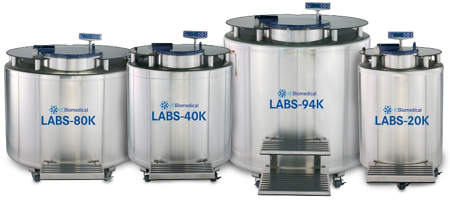
Bone Marrow Transplantation (Stem Cell Transplant)
0000-00-00


